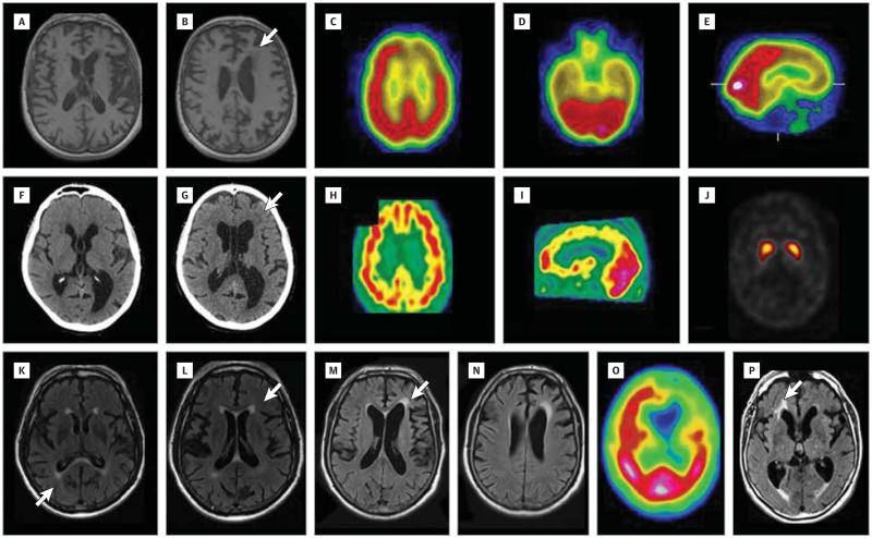
Figure 2. Brain Imaging of Patients Carrying SQSTM1 Mutations.
Axial T1-weighted magnetic resonance imaging (MRI) scans of the brain of proband 003 of family F297 reveal left-sided predominant frontal and temporal atrophy (A and B), a septum pellucidum cyst (A), and moderate periventricular hyposignals (B [arrow]). Technetium (Tc) 99m ethyl cysteinate dimer (ECD) single-photon emission computed tomographic (SPECT) scans on the axial (C and D) and sagittal (E) sections of proband 003 of family F297 reveal hypoperfusion of predominantly left frontal and bilateral temporal lobes. Computed tomographic scans of the brain of patient 005 of family F523 reveal moderate left-sided perisylvian (F) and bilateral frontal atrophy (G) associated with moderate white matter hypodensities (G [arrow]) and a septum pellucidum cyst (F and G). Tc 99m ECT-SPECT scans of the brain of patient 005 of family F523 reveal diffuse cerebral hypoperfusion on the axial (H) and sagittal (I) sections; the dopamine transporter (DaT) scan is normal (J). Axial fluid-attenuated inversion recovery (FLAIR) MRI scans of patient 010 of family F480 reveal predominantly right-sided perisylvian atrophy associated with moderate periventricular and callosal hypersignals (K and L [arrows]). Axial FLAIR MRI scans of patient 010 from family F1324 reveal predominantly left-sided frontal and perisylvian atrophy associated with moderate periventricular and callosal hypersignals (M [arrow] and N). Tc 99m ECT-SPECT scan of the brain of patient 010 from family F1324 reveals severe, predominantly left-sided frontal and temporal hypoperfusion (O). Axial FLAIR MRI scan of patient 005 from family FR1324 reveals bilateral frontal and temporal atrophy, with periventricular hypersignals (P [arrow]).

