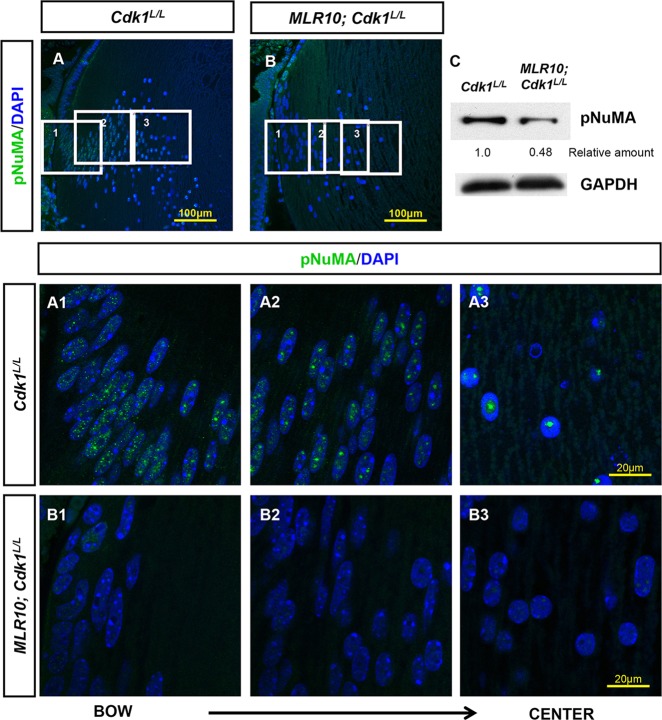
Fig. 5.
Lenses deficient in CDK1 failed to phosphorylate NuMA. (A-C) The monoclonal anti-phosphorylated threonine 2055 of NuMA (pNuMA) antibody detected the presence of pNuMA in Cdk1L/L lenses (A,C), and MLR10; Cdk1L/L lenses at E16.5 (B,C). Immunofluorescent (A,B) and western blot analysis (C) revealed reduced pNuMA in MLR10; Cdk1L/L lenses (B,C) relative to Cdk1L/L lenses (A,C). Three regions of Cdk1L/L (A, white boxes 1-3) and MLR10; Cdk1L/L (B, white boxes 1-3) lenses were selected for magnification (A1-3, B1-3, respectively). At the bow region of Cdk1L/L lenses, pNuMA diffusely spread across the entire nucleus (A1). As fiber cells mature towards the center of Cdk1L/L lenses, pNuMA localization appeared more punctate, finally converging on a single focus in the most mature fiber cell nuclei (A2,A3). By contrast, MLR10; Cdk1L/L lenses exhibit low levels of pNuMA post-mitotically, as both peripheral (B1) and central fibers (B3) lack pNuMA staining. Scale bars: 100 µm in A,B; 20 µm in A1-3,B1-3.

