Fig. 6.
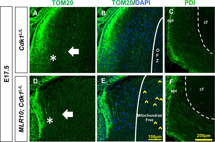
MLR10; Cdk1L/L lenses remove both mitochondria and endoplasmic reticulum, despite retaining nuclei. Mitochondria were detected by Tom20 (A,B,D,E) and endoplasmic reticulum by PDI (C,F) immunofluorescence (green staining) in E17.5 lenses. Cdk1L/L (A-C) and MLR10; Cdk1L/L (D-F) lens fiber cells lose their mitochondria (A,B,D,E) and endoplasmic reticulum (C,F) prior to reaching the center of the lens. Tom20 staining drops precipitously outside the most peripheral fiber cells (asterisks in A,D) but remains as punctate foci until the deep fiber cells of the central zone (arrows in A,D). Cdk1L/L lenses form an organelle-free zone (OFZ) lacking both mitochondria and nuclei (B). The MLR10; Cdk1L/L central lens fibers lack mitochondria, but retain nuclei (E, yellow arrowheads). Likewise, both control (C) and MLR10; Cdk1L/L (F) lenses remove PDI-staining endoplasmic reticulum from mature nuclear fiber cells (lack of green staining within the dotted line border in C,F). Nuclei were counterstained with DAPI (B,E). Scale bars: 100 µm A,B,D,E; 200 µm C,F. epi, lens epithelium; cf, central fiber cells.

