Figure 5.

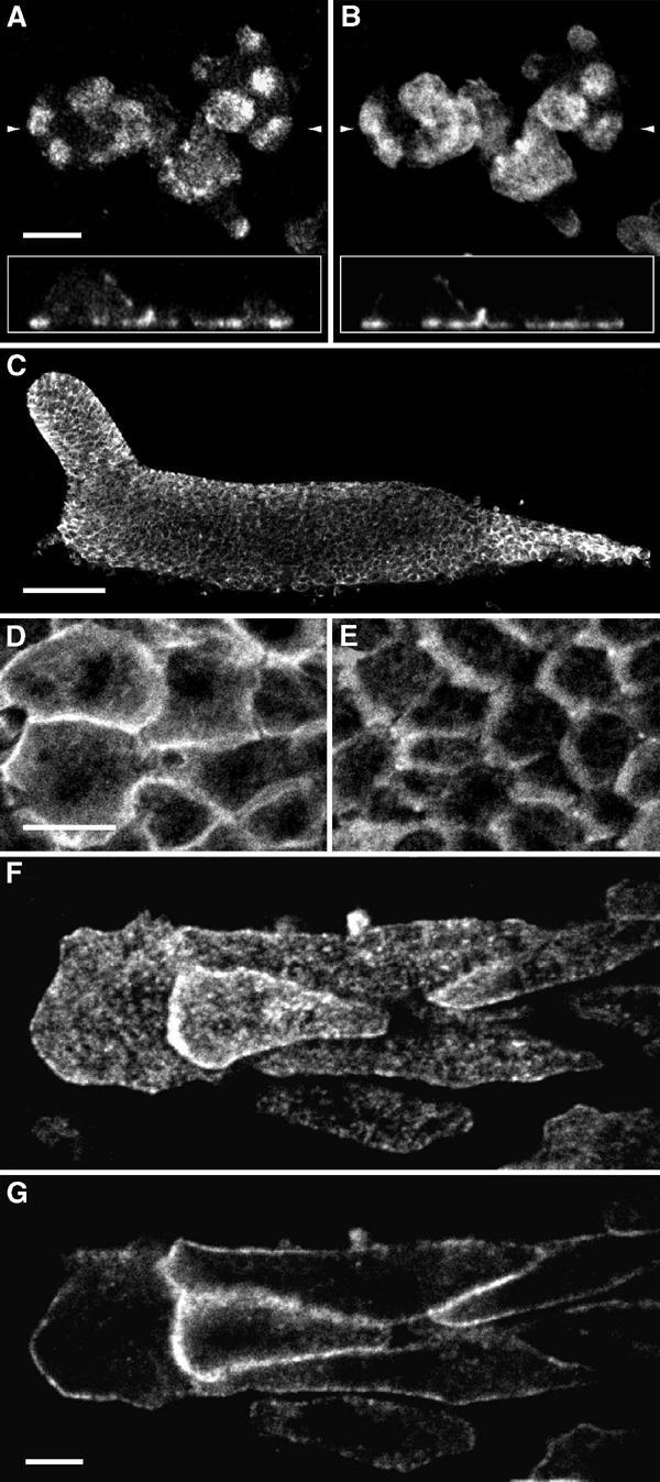
Localisation of talin B in cells at unicellular and multicellular stages of Ax2. (A, B) Cells at the growth phase were fixed with ethanol–formaldehyde and double-stained with the anti-talin B antibody (A) and fluorescent phalloidin (B). Focal plane immediately above the glass surface is shown. Insets show vertical sections reconstructed from horizontal sections made at intervals of 178 nm. Scale bar: 5 μm. The positions of the vertical sections are indicated by small arrows. (C) Whole-mount staining of a migrating slug with anti-talin B antibody. Anterior to the left. Scale bar: 50 μm. (D, E) Cells in the anterior prestalk region (D) and in the prespore region (E) at a higher magnification. Direction of cell movement is to the left. Scale bar: 10 μm. (F, G) Cells migrating away from a flattened slug. Focal planes right above the glass surface (F) and 500 nm above (G) are shown. Scale bar: 5 μm.
