
Figure 4.
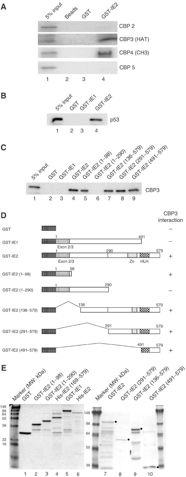
In vitro binding of IE2 and CBP. (A) Binding of GST-IE2 fusion proteins to in vitro-translated 35S-labeled CBP fragments. Beads (lane 2), GST beads (lane 3), or GST-IE2 beads (lane 4) were incubated with CBP2, CBP3, CBP4, or CBP5 fragments, then the bound complexes were separated by 10% SDS–PAGE and the gels subjected to autoradiography. (B) GST-IE2, but not GST-IE1, pulls down in vitro-translated p53. (C) Mapping of the CBP3-binding domain of IE2. Similar experiments were performed except that in vitro-translated 35S-methionine-labeled CBP3 was incubated with various GST-IE2 fragments. (D) Schematic diagram of the IE2 deletion mutants used in (C). (E) Purity of IE1 and IE2 derivatives used in Figures 3 and 4 shown by Coomassie blue staining. The molecular weight markers are indicated. The dots represent the expected position of the band corresponding to each protein.