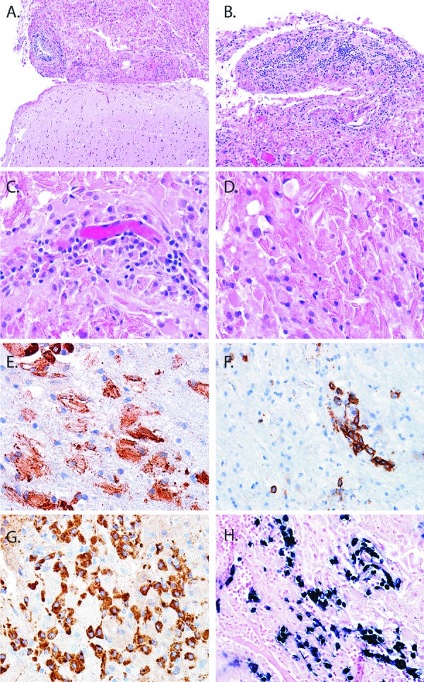
Figure 2. Pathology and immunophenotypic findings of CNS crystal-storing histiocytosis. A well circumscribed aggregate (top) was sharply demarcated from brain (bottom) (A). On higher magnification the infiltrate was composed of a mixed inflammatory infiltrate (B). A moderate number of plasma cells clustered around small blood vessels (C). Refractile, intracytoplasmic crystalline material was the hallmark feature of the lesion (D). Histiocytes expressed CD68 (E), while plasma cells expressed CD138 (F) and IgA by immunohistochemistry (G). In-situ hybridization studies for mRNA encoding immunoglobulin light chains showed κ-restriction (H).

