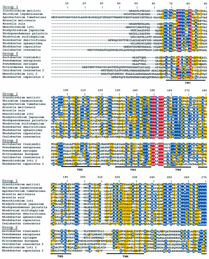
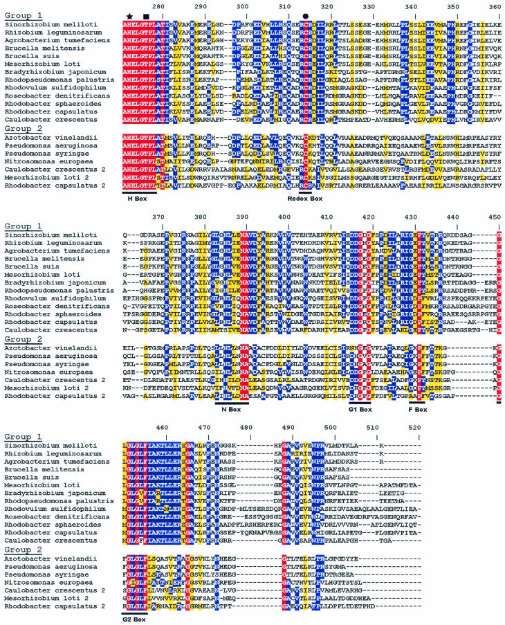
FIG. 2.
Alignment of RegB homologues present in genome databases. Membrane-spanning domains are indicated as regions TM1 through TM6. The site of histidine phosphorylation is denoted by a star within the H-box. The threonine residue important for phosphatase activity is indicated by a square. The location of the redox-active cysteine is denoted by a circle within the redox box. The ATP-binding domains are indicated as the N, G1, F, and G2 boxes. The color scheme is as described in the legend to Fig. 1.


