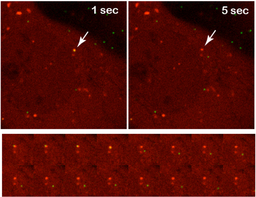
Figure 5. Adenovirus escaping from Gal-3 positive vesicular structures.
Top row) Detail of a single U2OS-FRT-Gal-3-mCherry cell. The arrow points to an Alexa488 labeled particle before (left) and after (right) escaping from the inside of a Gal-3 positive vesicular structure. Bottom row) Detailed image series of the endosomal escape process. Note that separation of the combined yellow signal into the green signal for the escaping particle and the red signal for the empty vesicular structure left behind.

