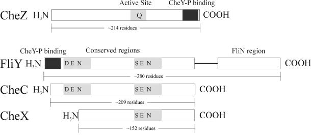
FIG. 8.
Schematic of CheY-P-hydrolyzing proteins. For CheZ, the C-terminal CheY-P binding region is shown in black and the area including what is thought to be the active site is shaded in gray. For FliY, the CheY-P binding site is shown in black. For FliY, CheC, and CheX, conserved regions are in gray, with highly conserved residues positioned as indicated.

