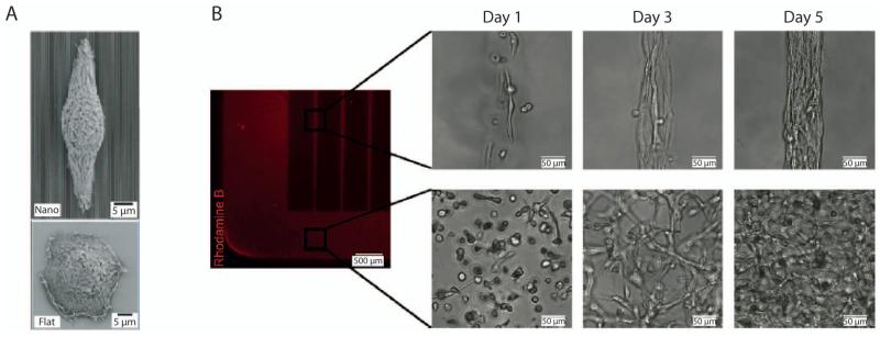
Figure 2.
Cell-Material Interactions. (A) Scanning electron microscopy images of a corneal epithelial cell on a nanograting topography (top) and flat surface (bottom).(Teixeira et al. 2003) Adapted with permission from Company of Biologists Ltd: [Journal of Cell Science], copyright (2003). (B) Fibroblast morphology and organization in patterned, 50μm-width rectangular (top) and unpatterned (bottom) gelatin methacrylate constructs.(Aubin et al. 2010) Adapted with permission from Elsevier: [Biomaterials], copyright (2010).

