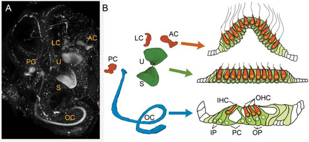
Figure 2.
The inner ear. A) Immunolabeling for Sox2 in an intact E15.5 inner ear shows the location of the sensory organs. The ear was cleared for confocal imaging using methyl salicylate and benzyl benzoate according to MacDonald and Rubel [81]. LC – lateral crista, AC– anterior crista, PC – posterior crista, U – utricle, S – saccule, OC – organ of Corti. B) A color coded model of the position of the Sox2-labeled sensory organs shown in A created by 3-dimensionally rendering tracings of the Sox2 regions in the individual confocal slices. In each of the inner ear organs, hair cells (orange) are arranged above the support cell layer (green). In the organ of Corti, the hair cells and support cells are highly specialized with obvious functional and morphological differences. In the vestibular system, these differences are not as pronounced. IHC – inner hair cell, OHC – outer hair cell, IP – inner phalangeal cell, PC – pillar cell, OP – outer phalangeal cell (Deiters’ cell).

