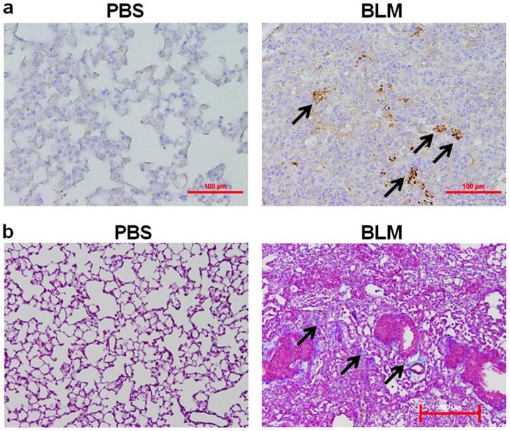
Figure 1.
Staining of β-catenin and collagen in lung tissue from mice with or without BLM challenge. Representative immunohistochemical staining for (a) β-catenin and (b) Masson’s trichrome staining for collagen in murine lung tissue isolated from mice with or without BLM challenge. Arrow shows the marked expression of (a) β-catenin (brown color) and (b) the deposition of collagen (blue color) in fibrotic foci. Scale bar, 200 μm.

