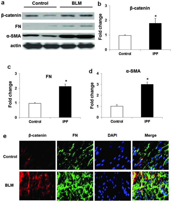
Figure 2.
Expression of β-catenin, α-SMA and FN in lung fibroblasts from mice with or without BLM challenge. (a) Representative western blot analysis (b–d) quantification of the expression of (b) β-catenin, (c) FN and (d) α-SMA) in lung fibroblasts from mice with or without BLM challenge. Data are expressed as means ± SEM of three independent experiments. *P<0.05 vs. cells from control mice. (e) Immunofloresent staining of β-catenin (red) and FN (green) in lung fibroblasts from mice with or without BLM challenge. Images were examined by immunofluorescence microscopy and recorded by using a 60× oil objective.

