Figure 2.

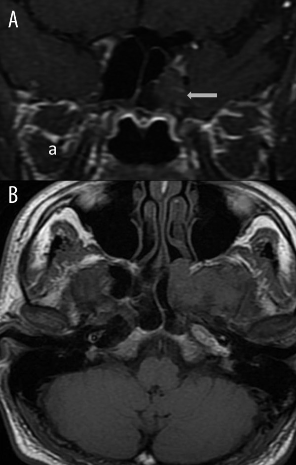
Contrast-enhanced T1W coronal (A) and axial (B) images: herniation of the temporal lobe into the sphenoid sinus is noticed (white arrows).

Contrast-enhanced T1W coronal (A) and axial (B) images: herniation of the temporal lobe into the sphenoid sinus is noticed (white arrows).