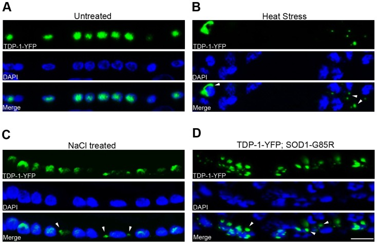
Figure 2. C. elegans TDP-1 forms cytoplasmic granules in neurons under proteotoxic stress.
Transgenic C. elegans expressing TDP-1-YFP under a neuronal promoter were treated with the indicated proteotoxic stressors prior to fixation, DAPI staining, and visualization by florescence microscopy. (A) In untreated C. elegans, TDP-1-YFP shows primarily nuclear localization. Ventral cord neurons are shown. (B) When heat-stressed at 28°C for 16 h, TDP-1-YFP shows cytoplasmic localization and forms granular structures. Nerve ring neurons are shown. (C) When treated with 0.4 M NaCl for 24 h, TDP-1-YFP also translocates to the cytoplasm and forms granules. Ventral cord neurons are shown. (D) When crossed to a transgenic strain stably expressing the ALS mutant SOD1-G85R in neurons, a subset of animals shows cytoplasmic translocation and granule formation of TDP-1-YFP. Arrowheads point to stress-induced TDP-1-YFP granules. Scale bar: 5 µM.

