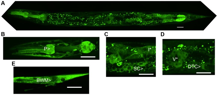
Figure 3. NATC-1 protein is expressed in many cells and tissues and localizes to the cytoplasm.
We generated transgenic natc-1(am138) animals expressing NATC-1::GFP fusion protein driven by the natc-1 promoter (WU1449). Confocal fluorescent microscope images of live animals are shown with head on the left and tail on the right. Green represents NATC-1::GFP fusion proteins, except for puncta in intestinal cells visible in panels A, C and D that reflect autofluorescent gut granules. (A) An image of an entire worm. NATC-1::GFP signal is visible in many cells and tissues throughout the animal, and the uniform staining pattern suggests cytoplasmic localization. (B–E) Higher magnification images display fluorescence in the pharynx (P>), sheath cells (SC>), intestinal cells (I*), distal tip cell (DTC>), vulva (V*) and body wall muscle (BWM>). Scale bar is 25 µm for all images.

