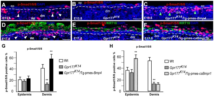
Figure 6. Transgenic pmes-Bmp4 reactivates Smad1/5/8 signaling in the dermal mesenchyme in Gpr177K14/Tg-pmes-Bmp4.
(A–F) Immunofluorescence detections for anti-phosphorylated-Smad1/5/8 (p-Smad1/5/8, red) on sections of autopods. P-Smad1/5/8 activity (white arrowheads) is preferentially decreased in dermis of limb skin in Gpr177K14 mice and increased in dermis of Gpr177K14/Tg-pmes-Bmp4 mice (green arrowheads) (A–C). Dash lines demarcate the border of epidermis and dermal mesenchyme. Immunofluorescence staining using antibodies against p-Smad1/5/8 (red) and KRT10 (green) on sections of dorsal autopod skin shows that p-Smad1/5/8 activity is only increased in epidermis of Gpr177K14/Tg-pmes-caBmpr-1a mice (D–F). b: basal layer; ep: epidermis; dm: dermis. Bars: 50 µm. (G–H). Quantification of pSmad1/5/8 positive cells in the epidermis and dermis of Gpr177K14/Tg-pmes-Bmp4 (G) and Gpr177K14/Tg-pmes-caBmpr-1a mice (H). Data are represented as mean ± SD. (**, P<0.01, n = 3–5).

