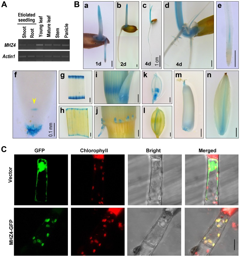
Figure 3. MHZ4 expression and protein subcellular localization.
(A) MHZ4 expression in different rice organs detected by RT-PCR. Actin1 was used as an internal control. (B) Tissue-specific expression of MHZ4 revealed by promoter-GUS analysis. Transgenic plants expressing MHZ4pro::GUS were used for analysis. Rice organs were stained for GUS for two days. At least 10 samples for each organ were observed and representative ones are presented. (a–c) 1 d- to 4 d-old etiolated seedlings. (d) GUS signals in adventitious roots and lateral roots of 4 d-old seedlings. (e) GUS staining in vascular tissues of root tips. (f) GUS staining in quiescent center (arrow head) and root caps of root tips. (g, h) GUS staining in segments of young (g) and mature (h) leaf blades. (i) GUS staining in young stem nodes and the base of axillary buds. (j) GUS staining in adventitious roots derived from nodes. (k) GUS staining in the anthers and pistils of young flowers. (l) GUS staining in the lemma of flowers. (m) Staining in the top and bottom of an ovary. (n) GUS staining in a developing grain. Bars are 1 mm except for those indicated. (C) Subcellular localization of MHZ4 in chloroplasts of tobacco glandular hairs as revealed by GFP-fusion protein. The constructs were transiently expressed in tobacco leaf cells by microprojectile bombardment. GFP fluorescence was detected using confocal microscopy. Red fluorescence indicates chlorophyll. Yellow color indicates co-localization of MHZ4 with chloroplasts. Bar = 10 µm.

