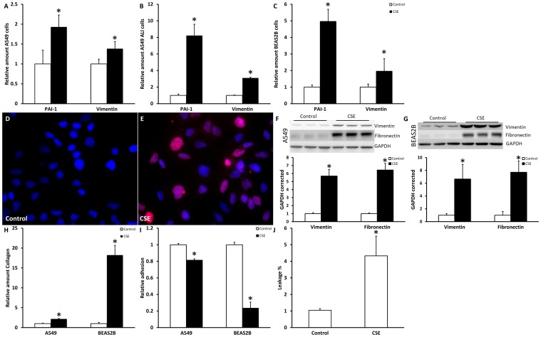
Figure 2. CSE increased mesenchymal markers in lung epithelial cells.
Cells were treated for 48 hours with respectively 2.5% CSE for A549 cells and 1.0% CSE for BEAS2B cells. mRNA levels of PAI-1 and vimentin were measured in A549 cell submerged (A), A549 cells at ALI (B) and BEAS2B cells (C) via qPCR. Data are expressed as mean+SD, * indicates p<0.05 compared to untreated controls. SNAIL staining of untreated control A549 cells (D) and A549 cells stimulated with CSE for 48 hours (E) as detected by immunofluorescence (red). Nuclei are counterstained using DAPI. Vimentin and fibronectin Western blot and quantification of untreated controls and 48 hours CSE treated A549 cells (F) and BEAS2B cells (G) in triplicate using GAPDH as a loading control. Collagen in the medium, measured using sircoll assay (H), Adhesion of A549 and BEAS2B cells (I) and leakage of A549 cells grown ALI after CSE exposure (J).

