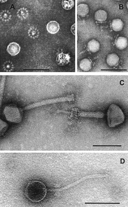
FIG. 2.
Morphological diversity of phages isolated from food fermentation. The negative-stain electron microscopic pictures show a Lactobacillus plantarum myovirus LP65 (note the contracted tail in the right phage in panel C), a L. plantarum siphovirus LP45 (panel D), and a Staphylococcus carnosus stc1 or -2 podovirus (panel B, side view of stc2; panel A, stc1 [from beneath, the phage baseplate becomes visible as a wheel-like structure]). All phages were isolated from meat (salami) fermentation. The bars are 100 nm.

