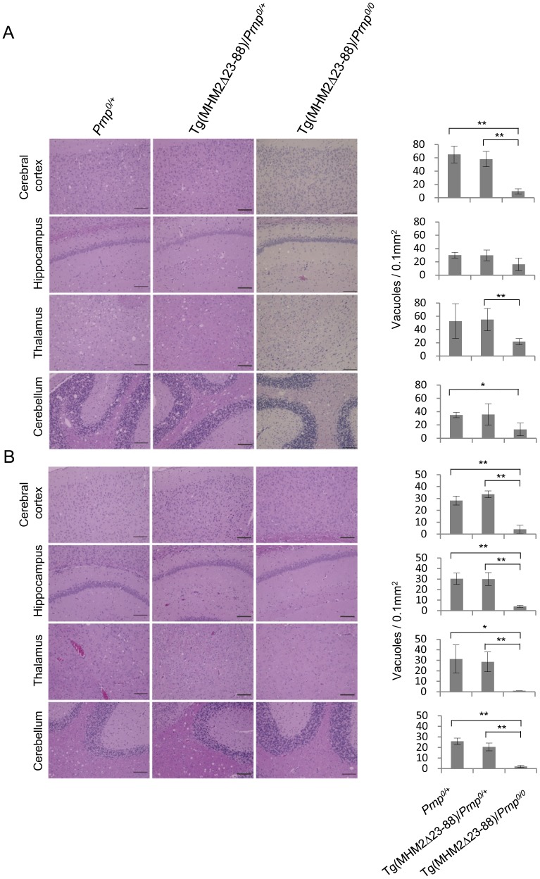
Figure 1. HE-stained brain sections from different genotypic mice inoculated with prions.
(A) Spongiosis is milder in the cerebral cortex, hippocampus, thalamus and cerebellum from 22L-inoculated, terminally ill Tg(MHM2Δ23-88)/Prnp0/0 mice than in 22L-inoculated, terminally ill Prnp0/+ and Tg(MHM2Δ23-88)/Prnp0/+ mice. (B) Spongiosis is observed in the cerebral cortex, hippocampus, thalamus and cerebellum from RML-inoculated, terminally ill Prnp0/+ and Tg(MHM2Δ23-88)/Prnp0/+ mice, but not from RML-inoculated, symptom-free Tg(MHM2Δ23-88)/Prnp0/0 mice. Vacuoles were counted in 0.1 mm2 in each brain area of different genotypic mice (n = 3–4/each genotype) and evaluated by Student's t-test. Scale bar, 100 µm. *, p<0.05; **, p<0.01.

