Figure 3.
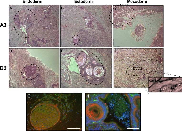
A3 and B2 ESC cell lines differentiate into cells from the three germ layers within teratomas.
Notes: Tumor assays of (A–C) A3-ESC exhibiting endoderm, ectoderm, and mesoderm, respectively. (D–F) B2-ESC exhibiting endoderm, ectoderm, and mesoderm, respectively. The selected A3-ESC images show (A) elongated columnar cells (acinar epithelium) with dark cytoplasm of the digestive organs. (B) The circular structure near the bottom is dermis sensory tissue consistent with the onion-like Pacinian corpuscle. (C) The circular structures closely resemble myofibrils of skeletal muscle. The B2-ESC developed (D) exocrine cells containing nuclei surrounded by small, cuboidal, uniformly dark stained cytoplasm. This is a defining characteristic of serous acini cells found in the secretory tissue. The B2-ESC tumors also developed (E) Pacinian corpuscles and (F) a region of cardiac muscle (dotted circle) with clearly defined sarcomere structures. Teratomas also display Tie-2 GFP+ and α-SMA RFP+ cells interacting with each other. (G) A3-ESC formed tissue resembling a lymph follicle with developing cells weakly expressing Tie-2 GFP+ surrounded by α-SMA RFP+ cells and strongly Tie-2 GFP+ cells that form venule-like structures. (H) B2-ESC form layers of α-SMA RFP+ cells that appear to be similar to the dermis sensory tissue of a pacinian corpuscle and are infiltrated with Tie-2 GFP+ cells that are indicative of small blood vessels. Scale bars =100 μm.
Abbreviations: α-SMA, alpha-smooth muscle actin; ESC, embryonic stem cells; GFP, green fluorescent protein; RFP, red fluorescent protein.

