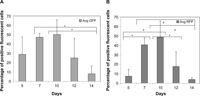
Figure 6.
Quantification of temporal expression of Tie-2 GFP+ and α-SMA RFP+ cells differentiating as monolayer cultures.
Notes: The Flk-1+ cells were re-plated as monolayers and allowed to differentiate overtime. The (A) Tie-2 GFP+ and (B) α-SMA RFP+ cells were then quantified as a function of time. Note that the first observed expression is seen at day 5, slightly earlier than the EB differentiation methods. *P<0.05 and #P<0.01, N≥3 distinct experiments.
Abbreviations: Avg, average; α-SMA, alpha-smooth muscle actin; GFP, green fluorescent protein; RFP, red fluorescent protein.

