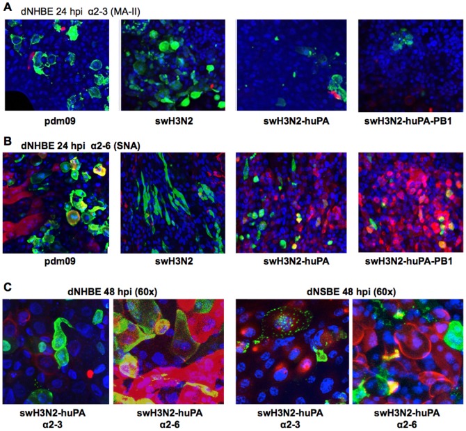
Figure 6. Immunofluorescence co-localization studies of virus and sialic acid expression on dNHBE cells.
To determine co-localization of virus with sialic acid expression, cells were examined for either (A) α2–3 sialic acid by MAA-II lectin stain (red) or (B) α2–6 SNA lectin stain (red). DAPI was used for nuclear staining (blue). Yellow indicates co-localization of the NP with sialic acids. (C) 60× magnification of swH3N2-huPA reassortant showed similar localization profiles as other viruses in dNHBE cells (left) and dNSBE cells [4] revealing minimal to no localization (yellow) in α2–3 stained cells versus more noticeable NP co-localization with α2–6 sialic acid.

