
Figure 3. Apigenin treatment does not affect hnRNP A1 or hnRNP A2 nucleocytoplasmic redistribution induced by EV71 infection. (A, B) Immunostaining study to determine hnRNP A1 and hnRNP A2 redistribution.
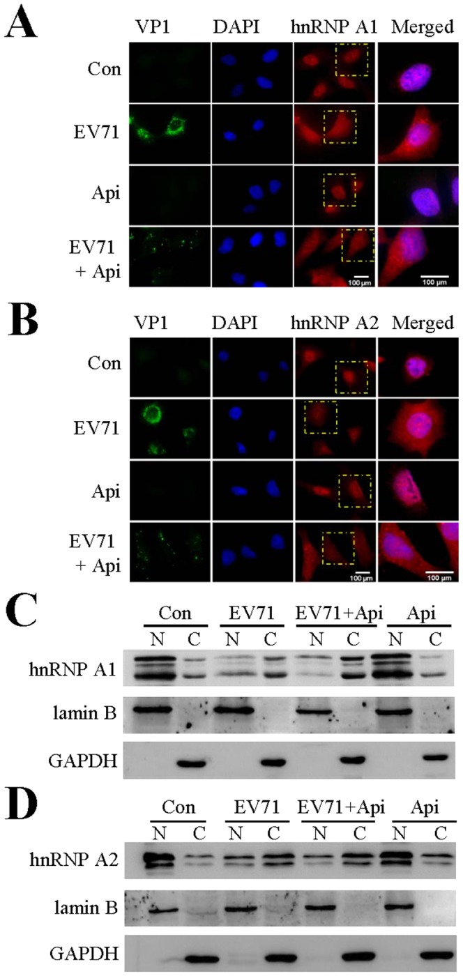
RD cells on coverslips were untreated or treated with 30 µM apigenin 2 hr prior to EV71 infection. The cells were infected with EV71 for 6 hr at an MOI of 40. The cells were then fixed and stained with rabbit anti-hnRNP A1 (A, red) or anti-hnRNP A2 (B, red), followed by Alexa Fluor 568-conjugated anti-rabbit antibody. EV71 VP1 protein expression was stained with a mouse anti-EV71 antibody (green) and the corresponding secondary antibody. The nuclei were visualized by staining with DAPI (blue). The merged images represent the areas within the yellow squares and highlight the relative location of hnRNP to the nuclei. (A) EV71 infection causes hnRNP A1 redistribution, while apigenin treatment does not affect hnRNP A1 redistribution. (B) EV71 infection causes hnRNP A2 redistribution and apigenin treatment does not affect hnRNP A2 redistribution. Images were collected with 400x magnifications and were processed using Image J. (C, D) hnRNP A1 and A2 redistribution by fractionation studies. Apigenin treated or control RD cells were uninfected or infected with EV71 at an MOI of 40 for 6 hr. The cells were harvested and fractionated for detection of cytosolic and nuclear proteins by immunoblotting. (C) redistribution of hnRNP A1 protein and (D) redistribution of hnRNP A2 protein. Lamin B and GAPDH were used as loading controls for nuclear and cytoplasmic proteins, respectively. Results are representatives of two independent experiments.