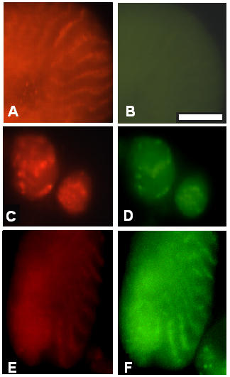
Figure 5.

The assembly of I-Z-I associated proteins is not affected in myosin heavy chain Mhc7 mutant. Immunofluorescence microscopy images representing the staining pattern obtained in Mhc7 mutants at 36–40 hours ASP and the 48 hours ASP. Pupae from the Mhc7 mutant stock were aged and after dissection, the tissues were spread and processed for immunofluorescence microscopy as described in Methods. All left panels are stained for projectin, and all right panels correspond to the double staining on the same cell for one of the other tested proteins. Panel A, C and E: projectin; panel B: myosin heavy chain; panels D and F: F-actin (FITC-labeled phalloidin). Panels C and D are at 36–40 hours ASP stage; panels A, B, E and F are at the 48 hours ASP stage. The image in panel B is purposefully overexposed to demonstrate the absence of myosin staining. Projectin forms Z-bands together with the other I-Z-I associated proteins, even in the complete absence of myosin. Scale bar in B is for panels A through F and represents 10 μm.
