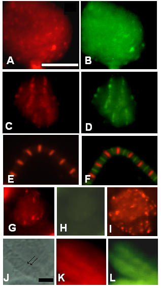
Figure 6.

The assembly of all I-Z-I associated proteins is affected in the KM88 actin mutant. Immunofluorescence microscopy images representing the staining pattern obtained in the E93K and KM88 actin mutants at 36–40 hours and 48 hours ASP as well as in purified adult myofibrils. Pupae from the E93K and KM88 mutant stocks were aged and after dissection, the tissues were spread and processed for immunofluorescence microscopy as described in Methods. Myofibrils were prepared from adult E93K and processed for immunofluorescence microscopy as described in Methods. Panels A through F represent data for the E93K mutant. Panels A and B: 36–40 hours ASP pupae stained respectively for projectin and F-actin; panels C and D: 48 hours ASP pupae stained respectively for projectin and F-actin; panels E and F: E93K adult myofibrils stained respectively for projectin and myosin + projectin. Panels G through L show data for the KM88 mutant. Panels G and H: 36–40 hours ASP pupae stained respectively for projectin and F-actin; panel I: 36–40 hours ASP pupae stained for kettin. Panel J-L: 60 hours ASP pupae, with J showing the phase contrast image and panels K and L stained respectively for projectin and kettin. Arrows in J indicate two visible Z-bands. Projectin forms Z-bands together with the other I-Z-I associated proteins in the E93K mutant, but not in the KM88 mutant pupae. Scale bar in A is for panels A through I and represents 10 μm. Scale bar in J is for panels J through L and represents 10 μm.
