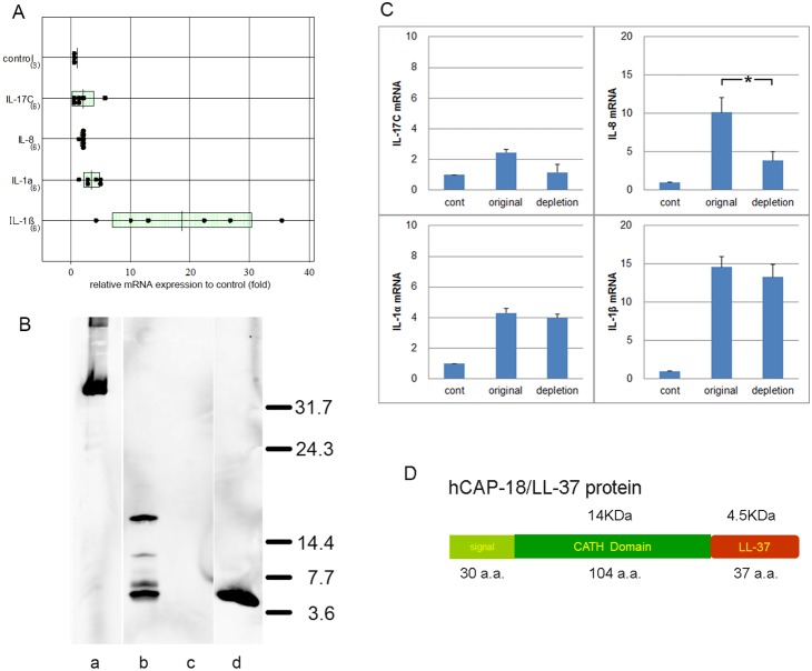
Figure 1. PPP vesicles stimulate expression of cytokine-encoding mRNAs in LSEs.
PPP vesicles were suspended in 1% (w/v) agar and mRNAs measured using qRT-PCR. A) All of IL-17C (2.00±1.79-fold), IL-8 (1.8±1.7-fold), IL-1α (3.47±1.28-fold), and IL-1β (18.67±11.72-fold) were upregulated compared to the levels in non-treated LSEs (controls). The levels of cathelicidin, IL-8, IL-1α, and IL-1β differed significantly from those in control sweat. B) Western blotting showed that the hCAP-18/LL-37-depleted PPP-VF sample contained bands equivalent to hCAP-18 (18 kDa; full-length); an intermediate-sized fragment (∼14 kDa); mature LL-37 (4.5 kDa); and two additional bands (lane b). Lane a: the GST-hCAP18 peptide prior to incubation; Lane b: PPP-VF before depletion; Lane c: PPP-VF after depletion of endogenous hCAP-18/LL-37; Lane d: synthetic LL-37 peptide (3.2 pmol). C) mRNA expression levels in LSEs stimulated by original and depleted PPP-VF, as calculated via qRT-PCR. *p<0.05. D) The illustration of structure of hCAP-18/LL-37.

