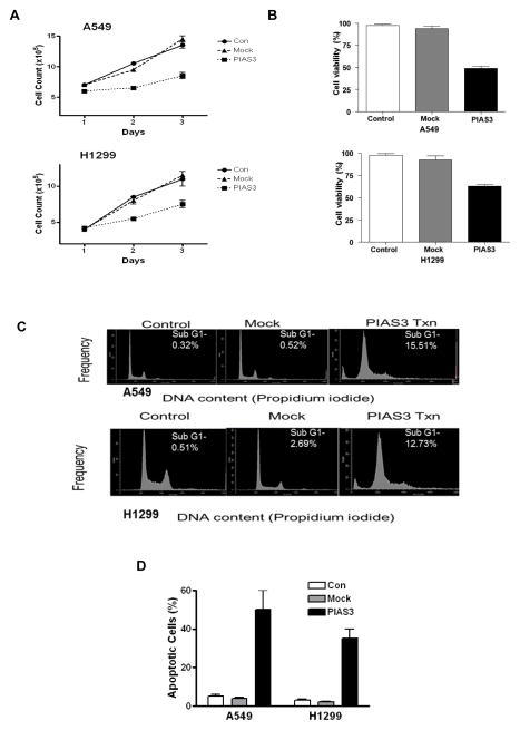
Figure 1. PIAS3 inhibits cell growth and induces apoptosis in non-small cell lung cancer cells.
A549 and H1299 cells were transfected with PIAS3 or empty plasmid (mock) or not transfected (control). In both cell lines, a significant decrease in proliferation was seen 24 h after the transfection by both cell count measurement (A) (p<0.01 for all groups) or MTS viability assay (B) (p<0.01 for all groups) compared to controls (N=4, data reported as mean ± SE). (C) Analysis of apoptosis by sub-G1 DNA content using flow cytometry of PIAS3 over-expressing cells. A549 and H1299 cell lines were transfected with empty plasmid or PIAS3. After 48 h, cells were stained by propidium iodide to analyze DNA content. The results shown are representative of at least three independent experiments. (D) Analysis of apoptosis by TUNEL staining in cells 48 h after transfection.

