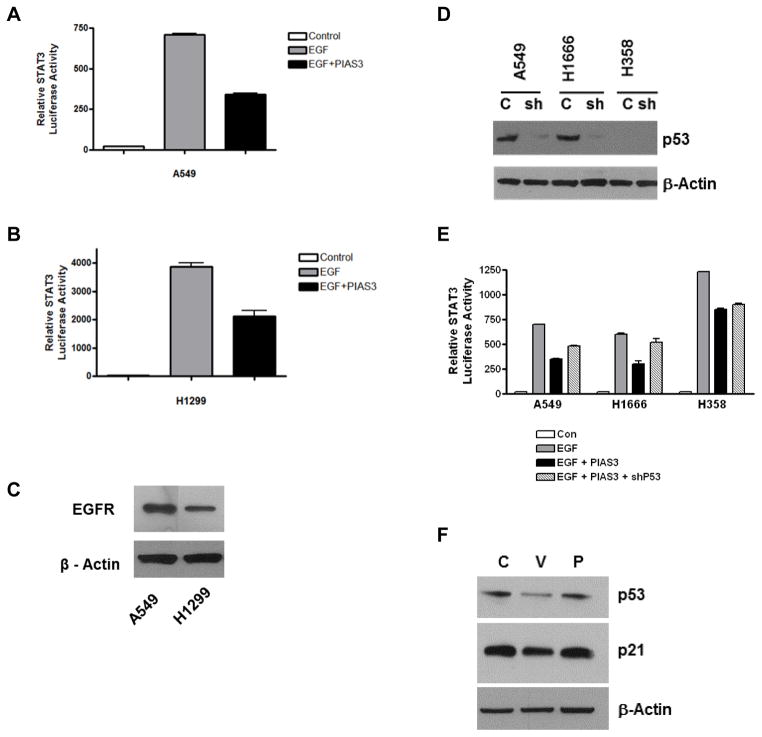
Figure 4. PIAS3 inhibits STAT3 activity independent of p53 status.
(A) A549 and (B) H1299 cells were transfected with STAT3 luciferase in the absence or presence of PIAS3. After 48 h, some cells were stimulated with EGF for 15 min after which lysates were prepared and analyzed. Results were normalized to protein concentration. (C) Western blot showing endogenous EGFR levels in A549 and H1299 cells. (D) Western blot of p53 levels. Protein lysates were prepared from A549, H1666 and H358 cells after 48 h incubation ± p53 shRNA lentivirus. (E) Combined effect of PIAS3 and p53 knockdown on EGF-stimulated STAT3 luciferase activity was done as described in Material and Methods. Each bar represents mean ± SD of three independent biological repeats. (F) Western blotting showing effects of PIAS3 transfection on p53 and p21 expression after 24 h in A549 cells.

