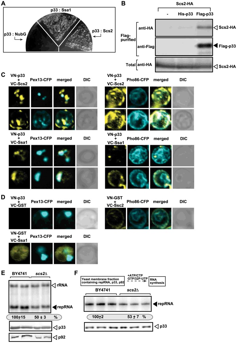
Figure 4. The tombusvirus p33 replication protein binds to the yeast Scs2p VAP protein in the ER.
(A) The split ubiquitin assay was used to test binding between p33 and Scs2p in wt (NMY51) yeast. The bait p33 was co-expressed with the shown prey proteins. SSA1 (HSP70 chaperone), and the empty prey vector (NubG) were used as positive and negative controls, respectively. (B) Co-purification of the Scs2p protein with the tombusvirus p33 replication protein. The FLAG-tagged p33 was purified from the membrane fractions of yeast extracts using a FLAG-affinity column. Top panel: Western blot analysis of co-purified 6xHA-tagged Scs2p using anti-HA antibody. Middle panel: Western blot of purified p33 (either His6- or Flag-tagged, as shown) detected with anti-FLAG antibody. Bottom panel: Western blot of 6xHA-tagged Scs2p in the total yeast extract using anti-HA antibody. (C) BiFC analysis of interactions between Scs2p and p33 and between Hsp70 (Ssa1p) and p33. Confocal laser microscopy images also show the peroxisomal localization of Pex13p marker protein (left panels) or Pho86-CFP ER-marker protein (right panels). The merged images at the top show the interaction between p33 and Scs2p and their partial co-localization with Pex13p-CFP or Pho86-CFP ER-marker, while the merged images at the bottom demonstrate the interaction between p33 and Ssa1 and their co-localization with Pex13p. DIC (differential interference contrast) images are shown on the right. Each row represents a separate yeast cell. Note that the Venus-(N-terminal portion) tag was fused to the N-terminus of p33 and the Venus-C tag was fused to host proteins, which are all N-terminal tags. Both p33 and Scs2p have cytosolic N-terminal regions. (D) Control BiFC experiments. Yeast was grown under similar conditions and images were taken as in panel C. (E) Decreased TBSV repRNA accumulation in scs2Δ yeast. To launch TBSV repRNA replication, we expressed His6-p33 and FLAG-tagged p92 from the copper-inducible CUP1 promoter and DI-72(+) repRNA from the galactose-inducible GAL1 promoter in the parental (BY4741) and in scs2Δ yeast strains. The yeast cells were cultured for 16 hours at 23°C on 2% galactose SC minimal media, and then for 24 h at 23°C on 2% galactose SC minimal media supplemented with 50 µM CuSO4. Northern blot analysis was used to detect DI-72(+) repRNA accumulation. The accumulation level of DI-72(+) repRNA was normalized based on 18S rRNA levels. Bottom panels: Western blot analysis of the accumulation level of His6-tagged p33 and FLAG-tagged p92 proteins using anti-His or anti-FLAG antibodies. Each experiment was performed three times. (F) Reduced activity of the tombusvirus replicase assembled in scs2Δ yeast. Top: Scheme of the experimental design. Denaturing PAGE analysis of in vitro replicase activity in the membrane-enriched fraction from wt and scs2Δ yeasts using the co-purified repRNA. The yeast cells were harvested for analysis at 24 h time point after launching TBSV replication. Note that this image shows the repRNAs made by the replicase in vitro. Each experiment was performed three times.

