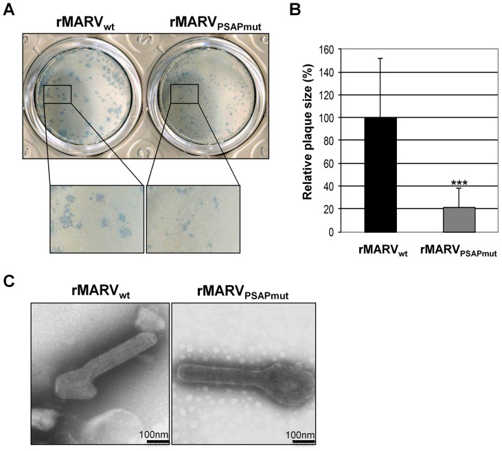
Figure 3. rMARVPSAPmut shows reduced plaque size but unaltered particle morphology.
(A) Vero E6 cells were infected with rMARVPSAPmut and rMARVwt and plaque formation monitored at 4 d p.i. by immunostaining. (B) Statistical analysis of the plaque size (n = 37). rMARVwt plaque size was set to 100%, p-value (***, P≤0.0001). (C) Morphology of viral particles. rMARVPSAPmut and rMARVwt viral particles were fixed, negatively stained with 2% phosphotungstic acid and analyzed by electron microscopy.

