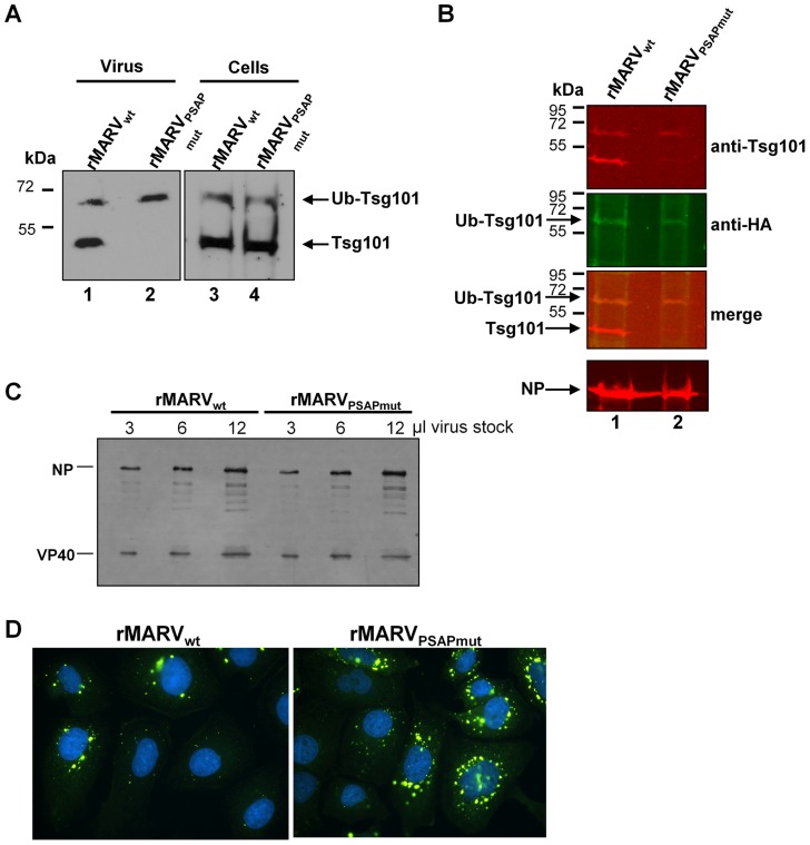
Figure 4. rMARVPSAPmut particles incorporate less Tsg101 and display similar infectivity.
(A) Tsg101 incorporation into MARV particles. Vero E6 cells were infected with rMARVwt or rMARVPSAPmut and virus particles released into the supernatant were pelleted through a 20% sucrose cushion at 48 h p.i. Virus pellets and cell lysates were subjected to SDS-PAGE and Western Blot analysis using Tsg101-specific antibody. (B) Detection of ubiquitinated form of Tsg101 in viral particles. Vero E6 cells were infected with rMARVwt or rMARVPSAPmut and subsequently transfected with HA-Ub expression plasmid. Virus particles were pelleted from the supernatants and analyzed by SDS-PAGE and Western Blot analysis using anti-Tsg101 and anti-HA specific primary antibodies and secondary antibodies for detection with the Odyssey imaging system (see merge image). (C, D) Comparison of virus infectivity. (C) Equal amounts of TCID50 units of rMARVPSAPmut and rMARVwt stock viruses were pelleted through 20% sucrose cushion, separated by SDS-PAGE and analyzed by Western Blot using NP- and VP40-specific antibodies. (D) Huh-7 cells grown on glass cover slips were inoculated with rMARVPSAPmut and rMARVwt normalized to nucleoprotein amount, fixed at 17 h p.i. and stained with DAPI and NP-specific antibody for detection of infected cells by immunofluorescence assay.

