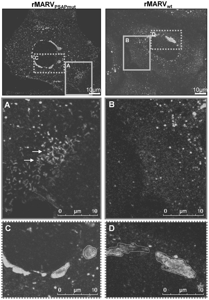
Figure 5. Infection with MARVPSAPmut results in compact inclusion bodies and accumulation of nucleocapsids in the periphery of cells.
Huh-7 cells were infected with rMARVwt or rMARVPSAPmut, fixed at 24 h p.i. and subjected to immunofluorescence staining using NP-specific antibodies. Samples were then analyzed by confocal laser scanning microscopy. Left panels: rMARVwt infection. Right panels: rMARVPSAPmut infection. Grey boxes in the upper pictures indicate different regions of the same cell that are shown in higher magnification below. (A) and (B) periphery of cells. (C) and (D) inclusion bodies. Arrows indicate nucleocapsids.

