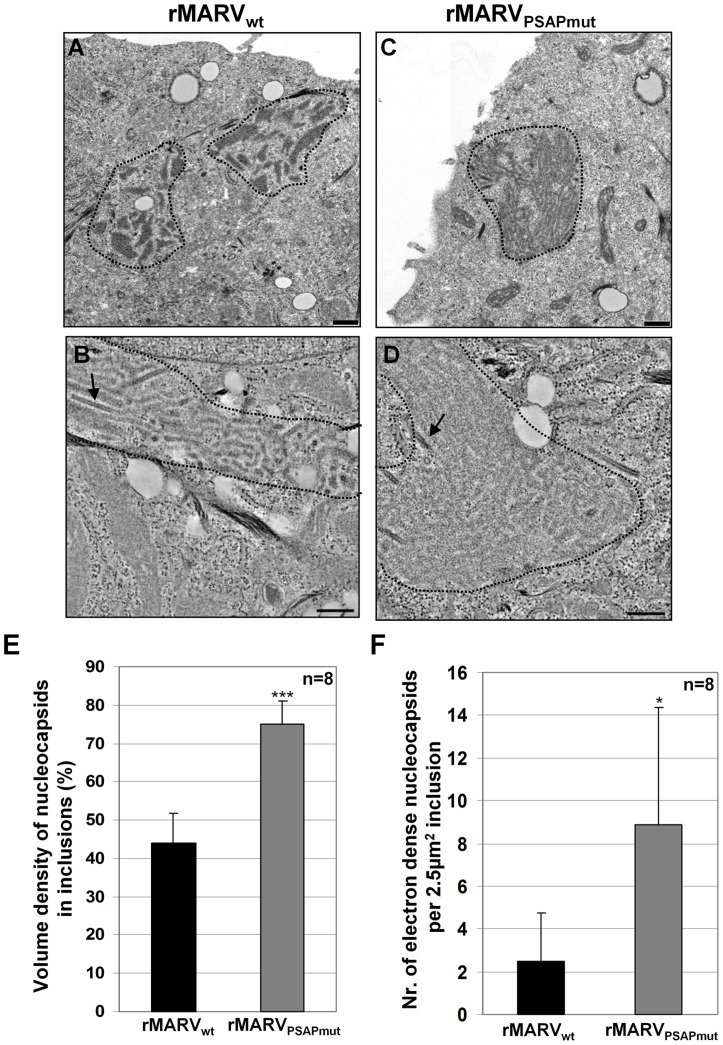
Figure 6. Inclusions in rMARVPSAPmut–infected cells are more densely packed with nucleocapsids than inclusions in rMARVwt–infected cells.
Huh-7 cells were infected with rMARVwt or rMARVPSAPmut. At 28 h p.i., cells were processed in two ways (i) fixed, scraped, pelleted and then embedded in Epoxy resin (A and C); or (ii) fixed and embedded in Epoxy resin on Thermanox slides (B and D). Ultrathin sections were stained with uranyl acetate and subjected to electron microscopy. (A–B) rMARVwt–infected cells, (C–D) rMARVPSAPmut–infected cells. Bars, 500 nm. (E) Morphometric analysis of inclusions. Volume density of nucleocapsids inside inclusions is shown, p-value (***, p≤0.0001). (F) Amount of electron dense (mature) nucleocapsids inside inclusions (see black arrows Fig. 6B und D) determined per 2.5 µm2 of inclusion at electron micrographs, p-value (*, p≤0.05).

