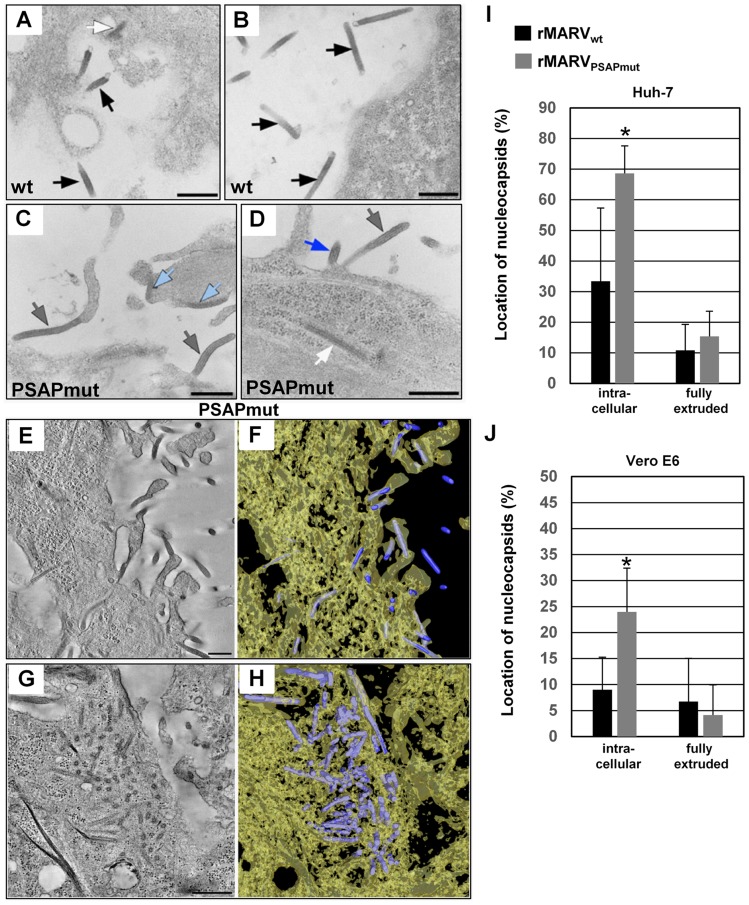
Figure 7. rMARVPSAPmut-infected cells contain more nucleocapsids in the cytoplasm and at early steps of budding than rMARVwt-infected cells.
Huh-7 and Vero E6 cells were infected with rMARVPSAPmut- or rMARVwt, fixed at 26 h p.i and embedded in epoxy resin. Cells were analyzed by thin section transmission electron microscopy (A–D) or electron tomography (E–J). (A–B) rMARVwt-infected Huh-7 cell displaying free virions (black arrows) and a nucleocapsid in the cytoplasm near the plasma membrane (white arrow in A). (C–D) rMARVPSAPmut-infected Huh-7 cells displaying fully protruded (grey arrows) or partially protruded (blue arrow in D) virus buds, and nucleocapsids bound to the plasma membrane (light blue arrows in C) or in the cytoplasm near the plasma membrane (white arrow in D). (E) 10 nm digital z-slice of an electron tomogram showing several nucleocapsids in the process of budding or in fully protruded virus buds in the periphery of rMARVPSAPmut-infected Huh-7 cell. (G) 9 nm digital z-slice of an electron tomogram showing accumulated nucleocapsids in the cytoplasm of rMARVPSAPmut-infected Huh-7 cell. (F, H) 3D surface representations of nucleocapsids (blue) and cytoplasm (yellow, semi-transparent) in the full tomograms for which z-slices are shown in Fig. 7E and 7G, respectively. Bars, 500 nm. (I–J) Quantification of the nucleocapsid distribution in tomograms from 300 nm thick sections of rMARVwt- or rMARVPSAPmut-infected Huh-7 or Vero E6 cells. Intracellular nucleocapsids (including cytoplasmic and those bound to plasma membrane, or partially extruded nucleocapsids) and fully extruded nucleocapsids were counted in a set (5 or more) of representative tomograms (p-value, *P≤0.05).

