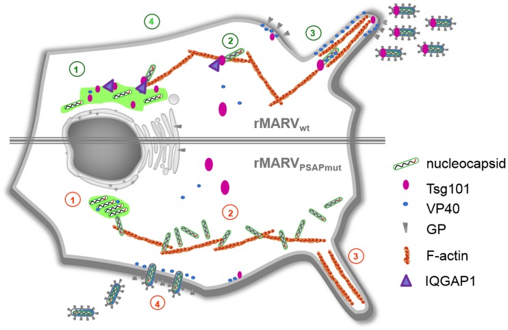
Figure 10. Schematic presentation of the rMARVPSAPmut phenotype in comparison to rMARVwt.
1. rMARVPSAPmut nucleocapsids accumulate in the inclusions which appear more dense and round. 2. The nucleocapsids accumulate in the cytosol and cell periphery. 3. rMARVwt virus buds preferentially from filopodia. 4. rMARVPSAPmut displays enhanced budding from the planar surface of cell. Numbers in red indicate the defective phenotype of MARVPSAPmut and numbers in green show rMARVwt infection.

