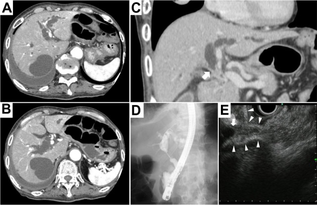
Figure 1.

Radiological images.
Notes: (A and B) Contrast-enhanced abdominal computed tomography revealed dilatation of the intra- and extrahepatic bile duct, including the common bile duct. (C) A computer-reconstructed coronal section image revealed obstruction of the distal common bile duct, which showed abrupt termination on the distal side. (D and E) Endoscopic retrograde cholangiopancreatography and endoscopic ultrasound revealed obstruction of the common bile duct secondary to the intraductal tumor. The arrow indicates the distal end of the common bile duct. The arrowheads indicate the wall of the common bile duct and cystic duct.
