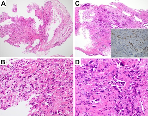
Figure 2.

Pathological specimens and cholangioscopic images.
Notes: (A and B) Adenocarcinoma cells in a specimen obtained by transbronchial biopsy. (A) ×40; (B) ×400. (C and D) Adenocarcinoma cells in a specimen obtained from the intraductal tumor. (C) ×40. Inset: thyroid transcription factor-1 stain. (D) ×400.
