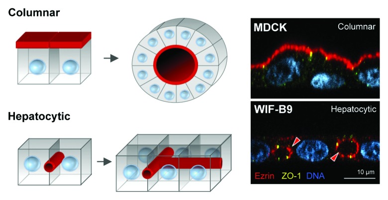
Figure 1. Columnar vs. hepatocytic polarity. Columnar epithelial cells form monolayers where multiple cells surround a central lumen (i.e., columnar polarity), whereas hepatocytes organize around tubular networks were the luminal domain is shared by no more than two cells (i.e., hepatocytic polarity), and each cell can have multiple luminal domains. Red arrowheads indicate the luminal domains marked by Ezrin. MDCK and WIF-B9 cells are kidney- and hepatocyte-derived culture models, respectively.
