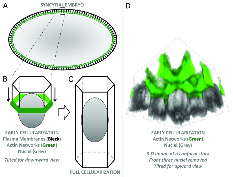
Figure 1. Plasma membrane organization of the syncytial Drosophila embryo. (A) Schematic cross-section of an early cellularization embryo. Black lines are plasma membranes. Green circles show actin networks forming a supracellular 3-D oval connecting all furrow tips around the embryo. (B) A 3-D schematic of a single hexagonal cell compartment containing a peripheral nucleus at early cellularization. Plasma membranes are invaginating from the surface with actin at the basal furrow tips. (C) A fully-formed cell. (D) 3-D imaging of F-actin (phalloidin staining) and nuclei (Nucleoporin 50-staining) of an early cellularization embryo using published materials and methods.7 The image is tilted for a view from the embryo interior toward the peripheral nuclei. The three front nuclei were deleted to allow a clear view of actin-coated plasma membranes and furrow tips.

An official website of the United States government
Here's how you know
Official websites use .gov
A
.gov website belongs to an official
government organization in the United States.
Secure .gov websites use HTTPS
A lock (
) or https:// means you've safely
connected to the .gov website. Share sensitive
information only on official, secure websites.
