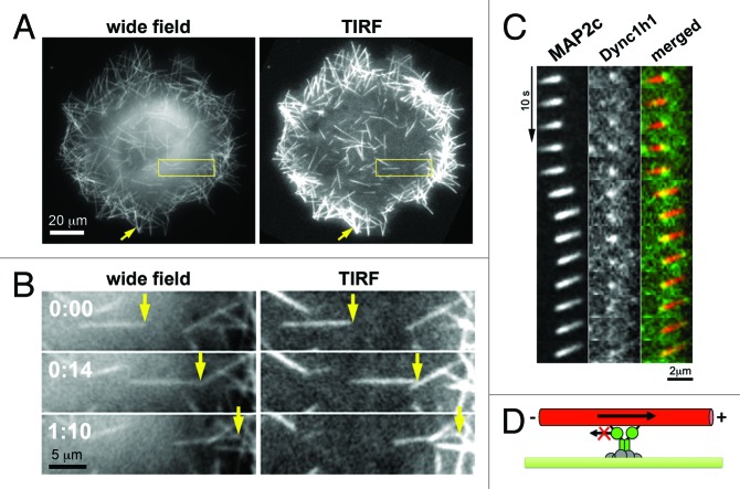
Figure 2. Direct observation of microtubule pushing by cortical dynein in living cells. (A) Images of COS7 cells transfected with the neuronal microtubule stabilizer MAP2c obtained shortly after nocodazole washout. In those conditions, short microtubules formed that were transported directionally within a distance up to ~200nm from the plasma membrane as observed by evanescent wave illumination in total internal refelction fluorescence (TIRF) microscopy. (B) Individual frames from video-microscopic observation of the boxed region in A. (C) Simultaneous observation of MAP2c-decorated short microtubules (left) and dynein heavy chain subunits (Dync1h1, middle) via TIRF microscopy. As expected for a direct, physical interaction between those components, the fluorescence signal derived from MAP2c-decorated microtubules is strongest at the localization of cortical dynein, suggesting that this microtubule region is closest to the plasma membrane and therefore excited most strongly by the evanescent wave of the TIRF microscope. (D) Schematic of the dynein-mediated microtubule pushing mechanism. This image was taken from26 (http://www.molbiolcell.org/content/25/1/95).

An official website of the United States government
Here's how you know
Official websites use .gov
A
.gov website belongs to an official
government organization in the United States.
Secure .gov websites use HTTPS
A lock (
) or https:// means you've safely
connected to the .gov website. Share sensitive
information only on official, secure websites.
