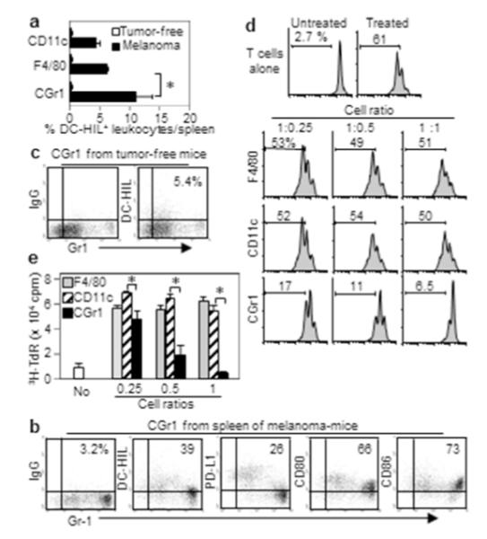
Figure 2. Melanoma induces DC-HIL expression by most potent CD11b+Gr1+ suppressors.

(a) Splenocytes from B16 melanoma-bearing or tumor-free mice (n=3) were assayed for % DCHIL+ cells in 3 myeloid populations. (b, c) CD11b+Gr1+ (CGr1) cells isolated from mice with (b) or without (c) melanoma were examined for expression of Gr1 and coinhibitory receptors (%). (d) These myeloid cells (increasing cell ratios) were cocultured with CFSE-labeled T-cells activated by anti-CD3/CD28 Ab. T-cell proliferation (%) was determined by FACS (histograms). (e) Purified myeloid cells were examined for T cell-stimulatory capacity. Different numbers of myeloid cells were pulsed with gp100 Ag and added to culture of CD8+ pmel-1 T-cells. Culture of T-cells with Ag served as control (No). T-cell proliferation was measured by 3H-thymidine (TdR) incorporation. *p<0.01.
