
Figure 3. DC-HIL mediates T cell-suppressor activity of CD11b+Gr1+ cells.
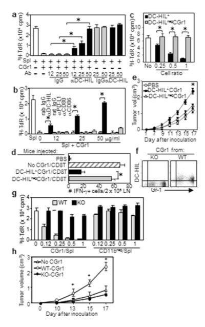
(a, b) CGr1 cells from melanoma-bearing mice were cocultured with pmel-1 splenocytes (Spl), gp100 Ag, and anti-DC-HIL mAb (a), anti-coinhibitor Ab or control IgG (b). 3H-TdR incorporation measured. (c) Undepleted (DC-HIL+) or DC-HIL-depleted (DC-HILneg) CGr1 (Supplementary method) were assayed for suppression of pmel-1 splenocyte proliferation triggered by Ag (increasing ratios). (d) Mice (n=5) injected with pmel-1 CD8+ T-cells and DC-HIL+CGr1 or DC-HILnegCGr1. Ten days after giving gp100, IFN-γ-producing cells in LN were counted. (e) Tumor growth following coinjection of DC-HIL+ or DC-HILneg CGr1 cells with B16 cells s.c. into naive mice (n=5). Using similar methods, DC-HIL−/−CGr1 cells were compared with DCHIL+/+ counterparts for DC-HIL expression by FACS (f), T-cell suppressing (g) and tumor-promoting ability (CD11bneg cells as control) (h). *p<0.01.