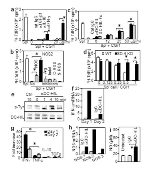
Figure 4. Crosslinked DC-HIL on CD11b+Gr1+ cells induces tyrosine phosphorylation and IFN-γ/iNOS expression.

(a-c) CD11b+Gr1+ (CGr1) cells cocultured with pmel-1 splenocytes (1:1 ratio) with inhibitors; including (a) anti-cytokine Ab; (b) 5 mM L-NG-monomethyl-arginine citrate (NOSs); 0.5 mM N6-(1-iminoethyl)-L-lysine (NOS-2); 1 mM N-hydroxyl-nor-arginine (Arg); 0.2 mM 1-methyl-tryptophan (Indol); 1000 U/ml catalase (C-ROS); and 200 U/ml superoxide dismutase (S-ROS); and (c) anti-DC-HIL mAb or DC-HIL-Fc. 3H-TdR uptake was measured. (d) CGr1 cells cocultured with SD-4+/+ or SD-4−/− pmel-1 splenocytes. (e-i) At varying times after crosslinking with Ab, CGr1 cells were assayed for: tyrosine-phosphorylation (p-Tyr) on DC-HIL protein (e); cytokine mRNA and secretion (f, g); mRNA of NOS genes (h); or NO production (i). Data (mean ± sd, n=3) are shown as fold increase relative to control. *p<0.01.
