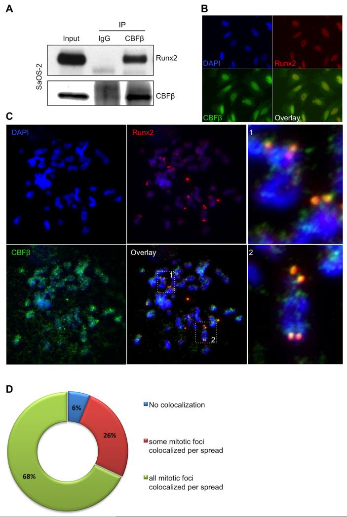
FIGURE 1. Analysis of CBFβ/RUNX2 colocalization during mitosis.
A, Immunoprecipitation of CBFβ was performed from asynchronous SaOS-2 whole cell lysates. A 22 KDa band corresponding to CBFβ (lower panel) was detected in the input (5%) and the CBFβ-IP samples, but not in those immunoprecipitated with the control, normal IgG. RUNX2 (60 KDa) was also seen only in the CBFβ-IP samples (Upper panel), indicating that is was co-immunoprecipitated with CBFβ and suggesting that RUNX2 and CBFβ remain as a complex. B, IF microscopy images of interphase SaOS-2 cells using antibodies for RUNX2 and CBFβ, and DAPI stain. Fluorescence data indicate that CBFβ is detected in both nuclei and cytoplasm, and RUNX2 is mainly located in the nucleus. In agreement with the IP results, co-localization of CBFβ with RUNX2 is observed (overlay image); C, IF of chromosomal spreads from SaOS-2 cells previously blocked in mitosis indicates that CBFβ is frequently located in chromosomes and co-localizes with RUNX2 in NORs (dotted squares 1 and 2 in overlay, enlarged on the right); D, Pie chart representing the distribution of co-localization of CBFβ with RUNX2 in mitotic spreads.

