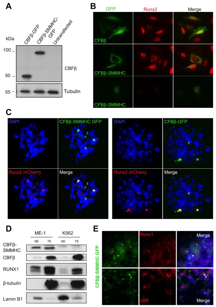
FIGURE 3. Leukemogenic CBFβ-SMMHC associates with NORs in mitotic chromosomes.
A, HeLa cells were transfected with CBFβ-GFP or CBFβ-SMMHC-GFP expression plasmids and western blot analysis from total cell extracts was performed. Detection with anti-CBFβ antibody shows proteins of sizes that correspond to exogenous (expression-plasmid-encoded) proteins (~54Kda and ~96 KDa, respectively); β-tubulin was used as a loading control. B, SaOS-2 cells transfected with CBFβ-GFP or CBFβ-SMMHC-GFP were fixed and labeled using an anti-RUNX2 antibody; GFP was detected by fluorescence only. CBFβ-SMMHC-GFP but not CBFβ-GFP caused cytoplasmic sequestration of RUNX2 as well as atypical distribution of RUNX2 in the nucleus; C, IF of chromosomal spreads from HeLa cells transfected with RUNX2-mCherry and CBFβ-GFP or CBFβ-SMMHC-GFP indicate that both normal and leukemic proteins remained associated with NORs during mitosis. D, Western blot of nuclear and total extracts from ME-1 and K562 leukemia cell lines to detect protein levels of CBFβ, CBFβ-SMMHC, and RUNX1; lamin B1 (nuclear) and β-tubulin (cytoplasmic) were used as loading and cell fractionation controls. E, Mitotic spreads from ME-1 cells expressing CBFβ-SMMHC-GFP were labeled with DAPI and examined via IF with antibodies to detect RUNX1 (upper panel) or UBF (lower panel).

