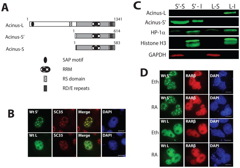
Figure 1. Acinus-L and Acinus-S′ have different sub-nuclear localization patterns.
A. Functional domains of the three human Acinus isoforms. B. The sub-nuclear localization of Acinus-L and Acinus-S′. C. Western blot analysis of Acinus-L and Acinus-S′ in soluble and insoluble fractions. D. RA does not affect the sub-nuclear localization of Acinus-L and Acinus-S′. In Panels B and D, COS7 cells were transfected with the indicated GFP-tagged wild type or mutant Acinus isoforms (B and D) and RFP-RARβ (D) expression vector DNAs. Twenty-four hr after transfection cells were fixed and proteins visualized using fluorescent microscopy (B) or treated with 10−6 M RA or Eth for an additional 5 hrs before fixation and visualization using fluorescent microscopy (D). Endogenous SC35 was labeled with anti-SC35 primary antibody and rhodamine conjugated secondary antibody (B). DAPI was used as a nuclear stain (B and D). Scale bar equals 10 μm (B and D). In Panel C, COS7 cells were transfected with wild type V5-Acinus-S′ (S′) or wild type V5-Acinus L (L) expression vector DNAs. Twenty-four hr after transfection soluble (S) and insoluble (I) fractions were prepared and analyzed by Western blot. Acinus-L and Acinus-S′ were detected using anti-V5 antibody. Histone H3 and HP1α were used as markers of the insoluble fraction and GAPDH was used as a marker of the soluble fraction.

