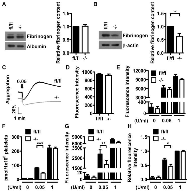
Figure 5.
Deficiency of platelet Dab2 causes a decrease in fibrinogen content and thrombin-induced ADP release and integrin αIIbβ3 activation. A, Platelet-poor-plasma (PPP) of fl/fl and −/− mice was collected and the fibrinogen content in PPP was determined by Western blotting using anti-fibrinogen antibody. The expression of albumin was used for the control of equal protein loading. The band intensity of fibrinogen normalized by albumin was quantified by ImageJ software. The data represent the mean ± SEM of 6 independent experiments. B, The platelets of fl/fl and −/− mice were collected and the fibrinogen contents were analyzed by Western blot using the anti-fibrinogen antibody. The expression of β-actin was used for the control of equal protein loading. The band intensity of fibrinogen normalized by β-actin was quantified by ImageJ software. The data represent the mean ± SEM of 4 independent experiments. *, p <0.05. C, 1 μg of fibrinogen was added to the fl/fl or −/− washed platelets from fl/fl or −/− mice followed by thrombin (0.05 U/ml) stimulation. Platelet aggregation was then recorded by a platelet aggregometer (Chrono-Log). Arrows indicate the point of agonist added. Essentially similar results were obtained in 3 independent experiments. D, The washed platelets from fl/fl and −/− mice were incubated with the FITC-conjugated anti-CD41 antibody and analyzed by flow cytometry. The data represent the mean ± SEM of 3 independent experiments E, Resting or thrombin-stimulated (0.05 U/ml and 1 U/ml) fl/fl and −/− platelets were incubated with the PE-conjugated anti-CD62P antibody and analyzed by flow cytometry. The corresponding isotype control antibody was used to define the background fluorescence signal. The data represent the mean ± SEM of 3–6 independent experiments. F, Supernatants from thrombin-stimulated fl/fl and −/− platelets were collected. ADP release assay was then performed and quantified using the GloMax 20/20 luminometer. The data represent the mean ± SEM of 5 independent experiments. ***, p < 0.001. G–H, Resting or thrombin-stimulated (0.05 U/ml and 1 U/ml) fl/fl and −/− platelets were incubated with the PE-conjugated JON/A antibody (panel G) or Alexa Fluor-488-conjugated fibrinogen (panel H) followed by flow cytometry. The corresponding isotype control antibody was used to define the background fluorescence signal. The data represent the mean ± SEM of 5–10 independent experiments. **, p < 0.01 and *, p < 0.05.

