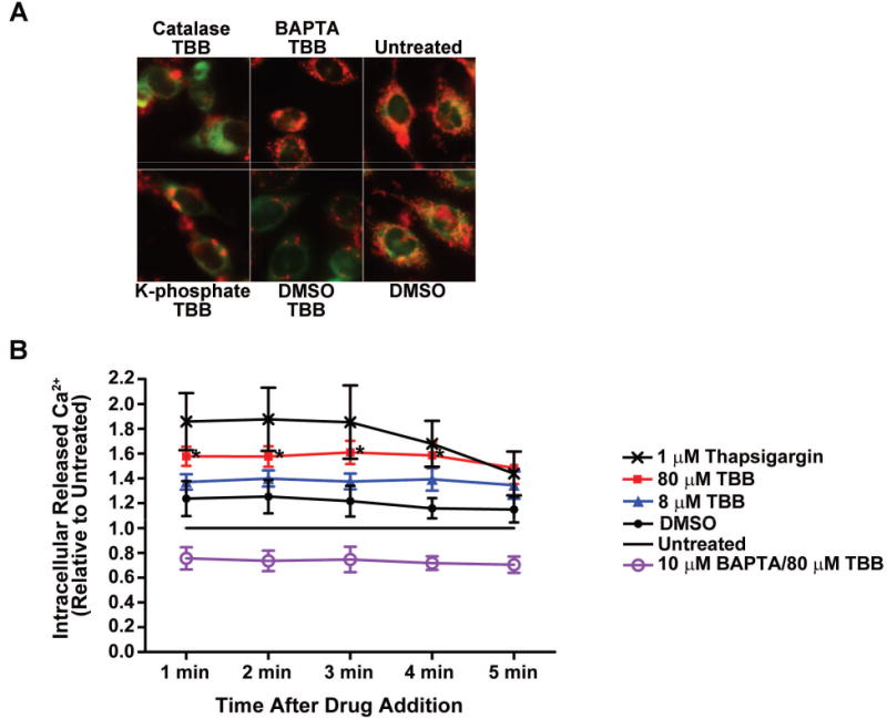
Fig. 3. Mitochondrial membrane potential change mediated by CK2 inhibition is blocked by pretreatment of cells with BAPTA but not catalase and is associated with transient release of intracellular Ca2+.

(A) PC3-LN4 cells were treated with catalase (3000 units) or BAPTA (10 μM) for 1 h followed by treatment with 80 μM TBB for 4 h. JC-1 loading was performed for the final 1 h of incubation. Controls included pretreatment for 1 h with drug solvent (DMSO for BAPTA; 50 mM potassium phosphate buffer pH 7.0 for catalase) followed by DMSO. Untreated cells were analyzed as a further control. Panels are accordingly labeled. Mitochondrial membrane potential was measured as described under materials and methods. (B) PC3-LN4 cells were loaded with FluoForte calcium assay dye, and with 10 μM BAPTA as indicated, for 1 h as described under Materials and Methods. Drug or diluent was added to each well as indicated in the legend (untreated cells received PBS diluent), and the plate immediately read once per min for 5 min. DMSO was used at the same concentration as 80 μM TBB. * p < 0.05 to untreated.
